How To Dapi Stain C Elegans Part 2 Imaging The Slide Information Center
Get comprehensive updates, key reports, and detailed insights compiled from verified editorial sources.
About to How To Dapi Stain C Elegans Part 2 Imaging The Slide

Here we use a fluorescence microscope to visualize and Learn how to use Fiji/ImageJ software to process a .czi or .tif microscopy Interns in the Microbial Ecology Laboratory at the Bermuda Institute of Ocean Sciences prepare A video from the Microbial Ecology Laboratory at the Bermuda Institute of Ocean Sciences (BIOS) demonstrating a protocol for ... This is a video teaching aid developed to supplement the text protocol This is Brush up on your coverslipping skills with these quick tips. Make sure you've chosen the right mounting medium first, though.
NOTE: Error in captions at 01:22. You must add 1mL green Get some quality control in your practice by making sure your
Key Details

Explore the key sources for How To Dapi Stain C Elegans Part 2 Imaging The Slide.
History

Stay updated on How To Dapi Stain C Elegans Part 2 Imaging The Slide's latest milestones.
Featured Video Reports & Highlights
Below is a handpicked selection of video coverage, expert reports, and highlights regarding How To Dapi Stain C Elegans Part 2 Imaging The Slide from verified contributors.

How to DAPI Stain C. elegans Part 2: Imaging the Slide
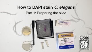
How to DAPI Stain C. elegans Part 1: Preparing the Slide
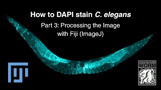
How to DAPI stain C. elegans Part 3: Processing the image with FIJI (ImageJ)

GFP Tagging & 4',6-Diamidino-2-phenylindole Staining-C. Elegans l Protocol Preview
Deep Dive
Data is compiled from public records and verified media reports.
Last Updated: May 22, 2026
Final Thoughts

For 2026, How To Dapi Stain C Elegans Part 2 Imaging The Slide remains one of the most searched-for profiles. Check back for the latest updates.
Disclaimer:


